Within BeAM a number of advanced microscopy techniques are available that are enabled by (scanning) transmission electron microscopes [(S)TEMs] and scanning probe microscopes [SPM]. For some techniques, there exists a dedicated instrument, and some techniques are available on multiple instruments, albeit with different parameters (e.g. spatial or spectral resolution).
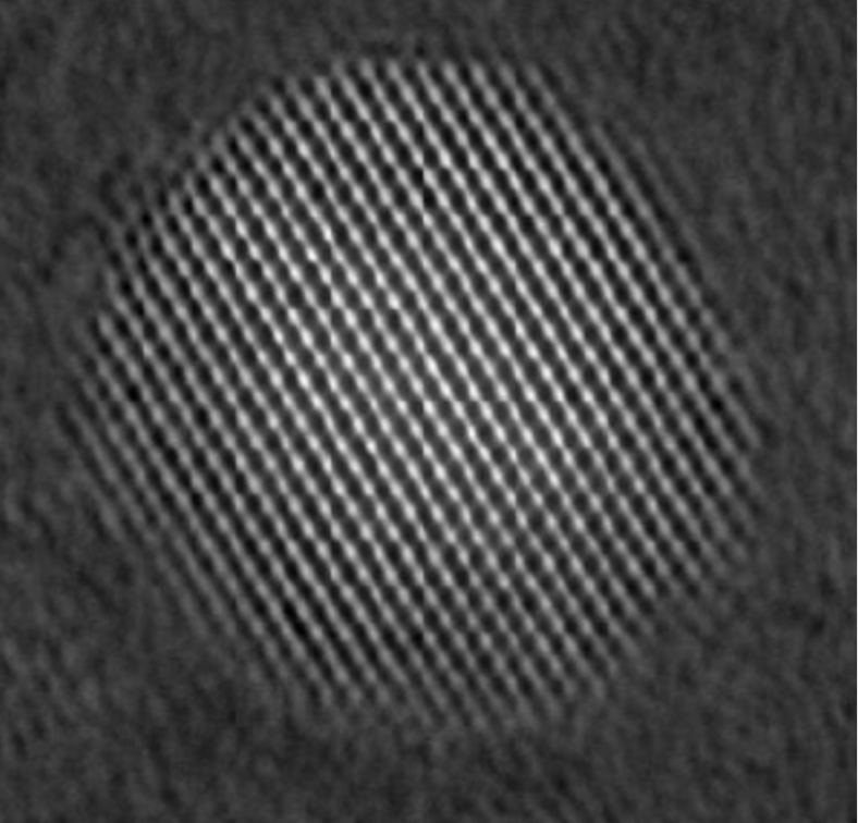
TEM Imaging illuminates the sample by a (nearly) plane electron wave and uses the microscope’s post-specimen optics to project an image onto the camera. A wide range of magnifications up to approx 106 is available, with the resolution reaching below 1 Angstrom. Further details …
Instruments: JEOL JEM2200FS, JEOL ARM200, JEOL JEM2100cryo, JEOL F200cryo, TFS Talos200
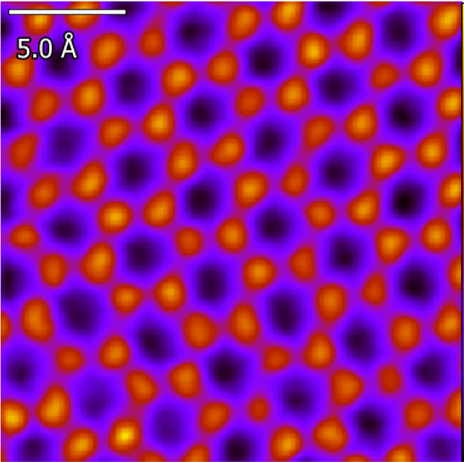
Scanning TEM techniques illuminates the sample with a focused electron beam that is scanned across the sample. A wide variety of signals can be acquired at each beam position, such as the number of transmitted electrons that impinge on a detector, X-rays generated by inelastic scattering, electron energy-loss spectrum (EELS), diffraction patterns, etc. Further details …
Instruments: JEOL JEM200, JEOL F200cryo, TFS Talos, TFS Titan, TFS SpectraUltra, Nion HERMES, Zeiss GeminiSEM500
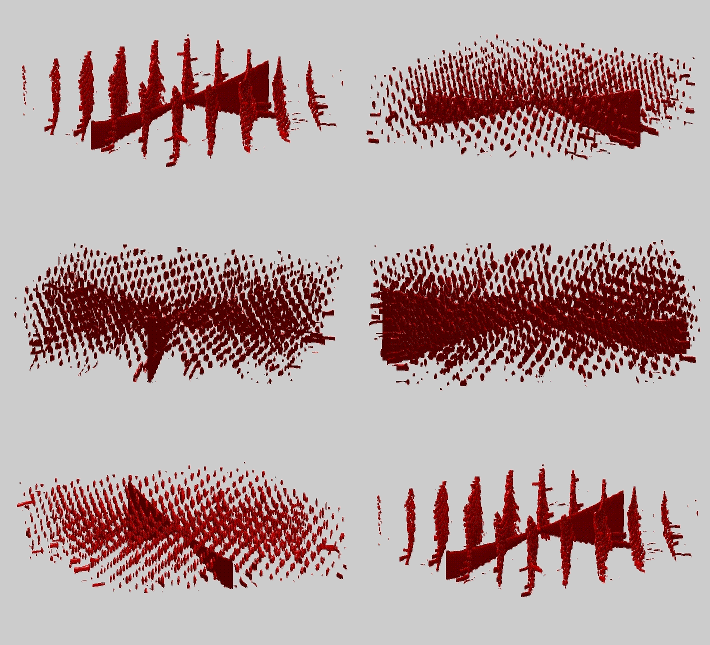
Electron diffraction records electrons scattered by the sample in reciprocal space. This measurement can be performed just once, or for many beam positions and provides information about crystallography, strain, radial distribution functions, etc. Further information …
Instruments: JEOL JEM200, JEOL F200cryo, TFS Talos, TFS Titan, TFS SpectraUltra, Nion HERMES, Zeiss GeminiSEM500
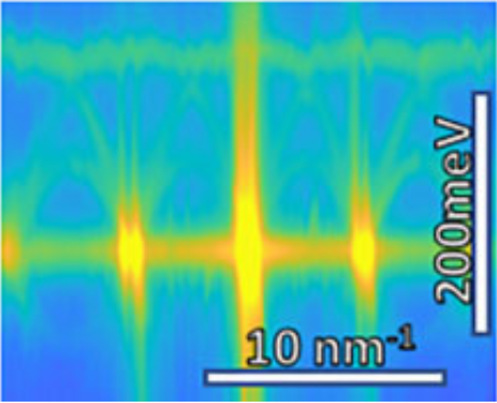
Electron Spectroscopy uses inelastic scattering of electrons by the sample, causing transmitted electrons to loose some of their energy (EELS) or gain energy by anihilating an excited state (typically phonons) of the sample (EEGS). Microscopes equipped with an EDX detetor can also detect characteristic X-rays that are generated when the electronic states relax after the excitation of an electron. Further information …
Instruments: Nion HERMES, TFS Spectra Ultra, JEL 2200FS, Talos,

